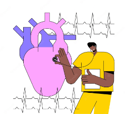
Diagnosis

Early and accurate diagnostic testing is a critical factor in detecting and optimizing coronary artery disease (CAD); thus, noninvasive cardiac imaging has become a central tool for CAD evaluation. The distinctive pathophysiology of atherosclerosis can be used together with imaging techniques to diagnose and assess the risk for CAD. Imaging modalities for cardiac risk stratification include various tools, such as noninvasive tests that visualize presymptomatic atherosclerosis and sophisticated radionuclide protocols that identify myocardial viability. (1)
- Electrocardiogram (ECG or EKG).This painless and quick test measures the electrical activity of the heart. It can show the speed at which your heart is beating.
- An Echocardiogram typically uses sound waves to create a picture of your heart. It can show how blood moves through the heart and heart valves.
- Heart (cardiac) CT scan. A heart CT scan can show calcium deposits and blockages in the heart arteries. Calcium deposits can narrow the arteries.
- Cardiac catheterization and angiogram. During cardiac catheterization, a cardiologist gently inserts a catheter into a blood vessel, usually in the wrist or groin. The catheter is gently guided to the heart. The dye will flow through the catheter, helps blood vessels show up better on the images, and outlines any blockages.
References:
- Miller, D. D., & Shaw, L. J. (2006). Coronary artery disease: diagnostic and prognostic models for reducing patient risk. The Journal of cardiovascular nursing, 21(6 Suppl 1), S2–S19. https://doi.org/10.1097/00005082-200611001-00002
- Image from https://www.freepik.com/premium-vector/heart-disease-diagnosis-abstract-concept-vector-illustration_32397379.htm

